Call us now :08045802326

 Send Inquiry
Ophthalmic Microscope
Price 140000 INR/ Unit
MOQ : 1 Unit
Ophthalmic Microscope Specification
- View Head
- Inclined, rotatable binocular
- Focus System
- Motorized fine/coarse adjustments
- Theory
- Ophthalmic Surgical Microscope designed for precision eye surgeries with optimal imaging quality.
- Drawtube
- Binocular
- Resolution
- 1920x1080 pixels
- Interface
- HDMI/USB 2.0
- Frame Rate
- 30 fps (live view)
- Focal Distance
- 200 mm Millimeter (mm)
- Magnification
- 5x, 8x, 12.5x, 16x, 25x (step magnification changer)
- Dimensions
- Base: 600 x 600 x 1500 mm (HxWxD) Millimeter (mm)
- Focus Range
- 50 mm motorized Millimeter (mm)
- Eyepieces
- 12.5x widefield
- Eyepiece Tube
- Inclined 45, 360 rotatable
- Illumination
- Coaxial LED with adjustable intensity
- Coarse Adjustment Range
- 35 mm
- Fine Adjustment Range
- 5 mm
- Still Image Capture Resolution
- up to 1920x1080 pixels
- Video Capture Resolution
- Full HD 1080p
- Image Format
- JPEG, BMP, DICOM
- Objective Achromatic
- f=200 mm (standard), interchangeable
- Light Source
- LED 50,000 hours life
About Ophthalmic Microscope
Since our inception in 1985, we have been counted amongst the leading manufacturers, exporters and suppliers of Ophthalmic Microscope. This microscope is basically used for performing posterior and anterior segment ophthalmic surgeries of human eye. This microscope is manufactured with high level of precision by our experienced professionals using pristine quality components with the aid of innovative techniques. Apart from this, our honorable patrons can avail this Ophthalmic Microscope in several technical specifications at the lowest prices from us.
Features:
- Longer service life
- Easy to operate
- Accurate result
- Compact design
Precision Imaging for Critical Eye Surgeries
This ophthalmic surgical microscope delivers exceptional clarity for complex eye operations. Its achromatic objective lens minimises distortion, while multiple step magnifications (from 5x up to 25x) allow surgeons to customise views based on procedural needs. The 1920x1080 pixel HD camera output further enhances diagnostic ability and documentation.
Ergonomic and User-Friendly Design
With an inclined, rotatable binocular head and a 45 eyepiece tube, this microscope supports comfortable use over extended surgeries. The motorized focus and coarse/fine adjustment options enable seamless, precise focusing. Easy connectivity using HDMI and USB 2.0 simplifies data output and workflow integration within any clinical setup.
FAQs of Ophthalmic Microscope:
Q: How does the step magnification changer benefit ophthalmic procedures?
A: The step magnification changer provides selectable magnification levels (5x, 8x, 12.5x, 16x, 25x), allowing surgeons to switch between detailed close-ups or broader views as required during delicate eye surgeries, thereby improving surgical flexibility and precision.Q: What image formats and output interfaces are supported for documentation?
A: The microscope supports JPEG, BMP, and DICOM formats for still images, and delivers video in full HD 1080p at 30 frames per second. HDMI and USB 2.0 interfaces enable seamless integration with external monitors, recorders, or computers.Q: When should the motorized focus system be used during surgery?
A: The motorized focus system should be used when fine-tuning or quick depth adjustments are needed. Surgeons can utilize the 50mm motorized range and 5mm fine adjustment to maintain optimal clarity as intraocular structures or surgical instruments shift during operations.Q: Where is the microscope best suited for installation?
A: The device is ideal for installation in ophthalmic surgical theatres or procedures rooms where precision eye surgeries are performed. Its ergonomic design and base dimensions (600 x 600 x 1500 mm) ensure stability and adaptability to standard clinical environments.Q: What is the expected lifespan of the microscopes LED illumination?
A: The built-in coaxial LED light source boasts a remarkable lifespan of approximately 50,000 hours, ensuring long-term, dependable use without frequent bulb replacements, thus reducing ongoing maintenance costs.Q: How can captured images and videos be utilised for patient care?
A: Captured images (JPEG, BMP, DICOM) and full HD videos can be instantly stored, reviewed, and shared for patient records, post-operative analysis, or case presentations, enhancing both patient documentation and collaborative care processes.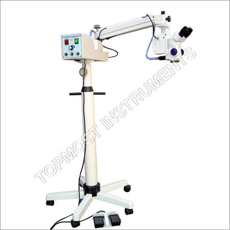
Tell us about your requirement

Price:
Quantity
Select Unit
- 50
- 100
- 200
- 250
- 500
- 1000+
Additional detail
Mobile number
Email

More Products in Opthalmic Microscope Category
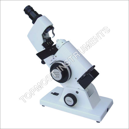
Lensometer .
Price 25000 INR / Unit
Minimum Order Quantity : 1 Piece
Voltage : 220440 Volt (v)
Color : White
Usage : Hospital
Material : MS
Our Products

4284, Behind B.D. School, Cross Road 2, Ambala Cantt - 133001, Haryana, India

Mr. G. S. DHIMAN
(General Manager)
Mobile :08045802326
Mr. B. S. Dhiman
 Send Inquiry
 Send SMS
 Call Me Free
Developed and Managed by Infocom Network Private Limited.
